
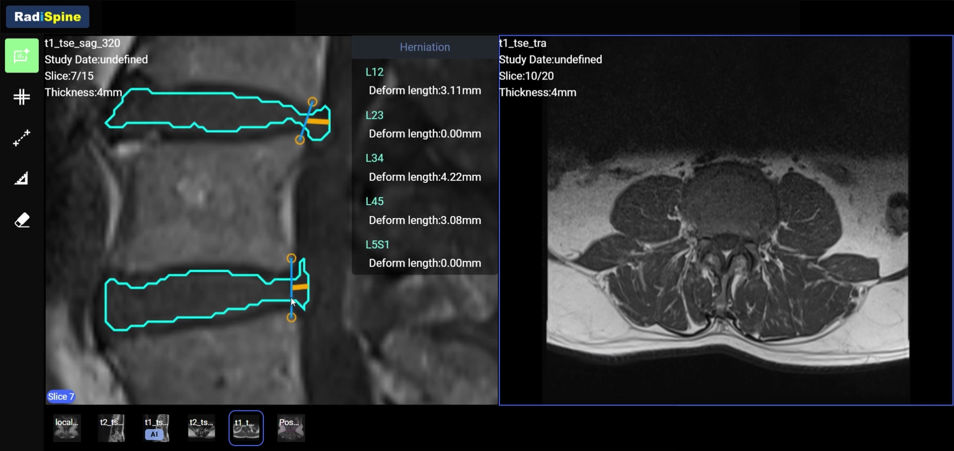
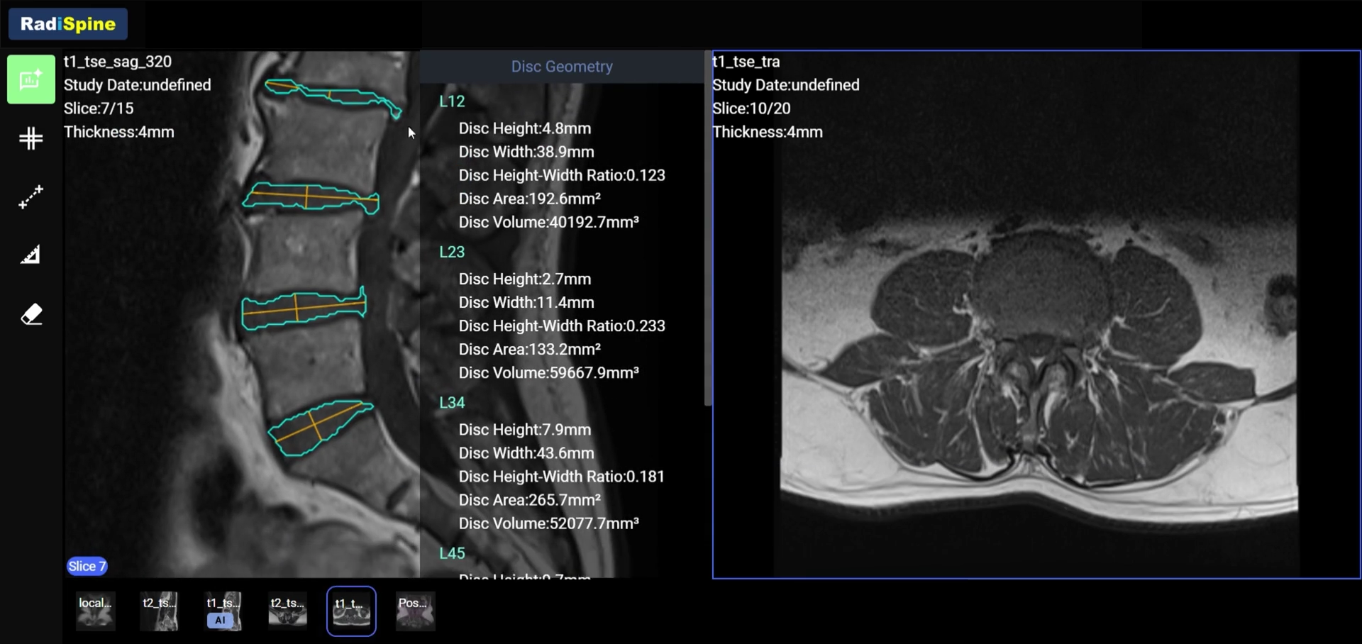
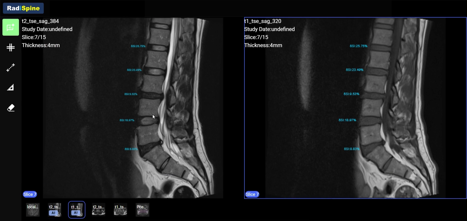
RadiSpine

AI reveals image detail. Supporting clinical judgment.

FOUR CORE ANALYSES

Four core functions

SPINAL STENOSIS

Make compression measurable.

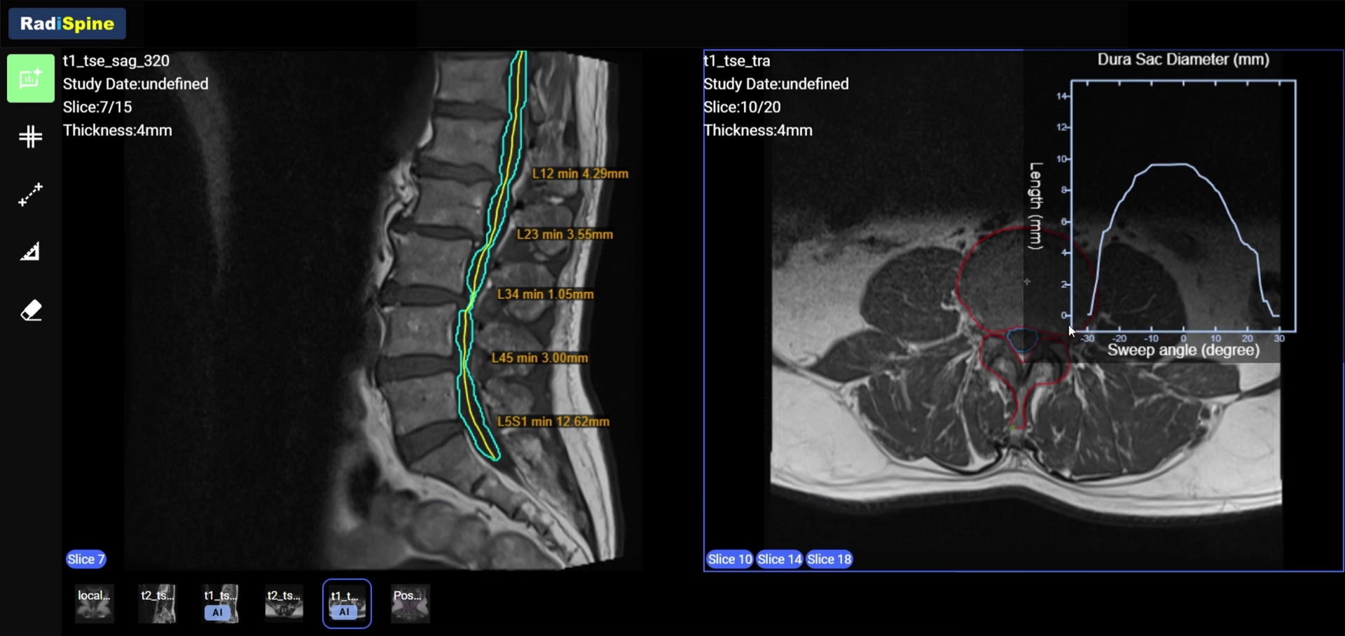
AI segments the dural sac and measures its diameter to show differences across lumbar levels. Comparing the original images with the measurements helps physicians identify the location and extent of narrowing and assess compression.

  • Dural sac segmentation
  • Diameter measurements and comparison across levels
  • Stenosis features alongside the original images
Spinal stenosis analysis

From images to insights. Explore RadiSpine.

RadiSpine workstation demonstration

BUILT AROUND YOUR WORKFLOW

AI-assisted analysis. Efficiency in clinical practice.

Connect image import, AI analysis and physician review, bringing quantitative findings into interpretation and reporting at each step of the clinical workflow.

RadiSpineINPUT

REGULATORY & CLINICAL EVIDENCE

Evidence builds trust. Innovation advances care.

Clinical research and adherence to medical device regulations and standards provide evidence for analytical accuracy and a foundation for trusted clinical innovation.

Product regulatory information

TFDA · TAIWAN

Taiwan Class II medical device

Taiwan Class II medical device clearance for the AI-based quantitative spine MRI analysis system.

Taiwan medical device manufacturing license No. 008564

CE · EUROPE

EU medical device conformity marking

CE marking provides a regulatory basis for use in the European Union market.

European Union | CE

Clinical research & publications

CLINICAL STUDY

Taipei Veterans General Hospital | IRB approval

The functional validation study received IRB approval and institutional authorization from Taipei Veterans General Hospital, enabling clinical validation under research ethics requirements.

IRB: 2024-08-008C · 12 Sep 2024

PUBLICATION

Neurospine | Published research

Research on automated lumbar MRI segmentation and quantitative assessment was published in Neurospine in 2024, presenting the technical methods and analysis performance in a peer-reviewed journal.

Read the research paper
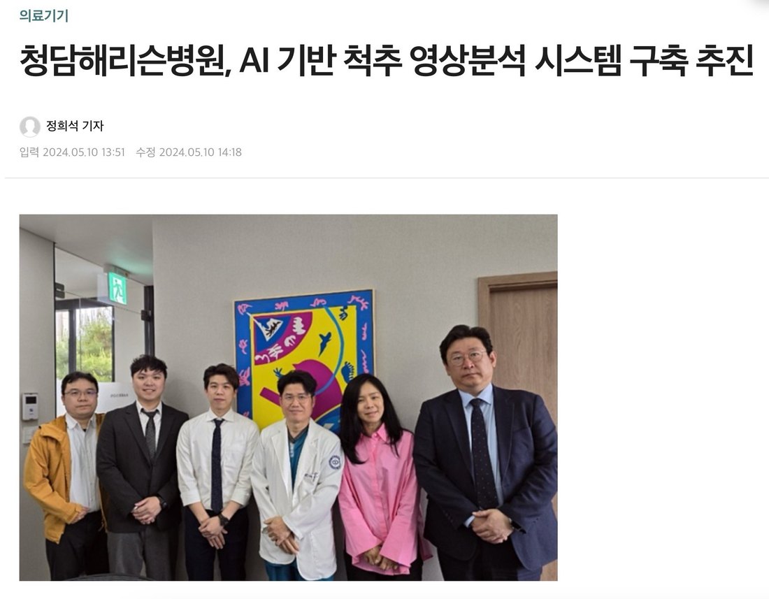
Group photograph of the Harrison Spinartus Hospital collaboration team

Clinical implementation | Seoul, South Korea

Harrison Spinartus Hospital

Working with Dr. Kim Hyun-sung’s team, we deployed an in-hospital AI imaging analysis service to support pre- and postoperative lumbar assessment. Quantitative information complements clinical interpretation and extends the role of imaging analysis in patient care.

Research meeting with CHA University and CHA Bundang Medical Center

Research collaboration | Seongnam, South Korea

CHA University / CHA Bundang Medical Center

In collaboration with Dr. Han In-bum’s team, we are researching AI-based quantitative assessment of recovery after lumbar fusion. By combining medical imaging analysis with clinical research needs, the project explores the potential of quantitative imaging indicators in postoperative assessment.

Medical device clearance, research ethics approval and journal publication each have a distinct scope. Product use is governed by the approved labeling.

THE NEXT CHAPTER

The 2.0 roadmap: multimodal imaging analysis

Further integrate multimodal imaging analysis and smart reporting, bringing richer quantitative information to clinical interpretation and conversations with patients.

REPORT

Smart Report

Further integrate and visualize analysis results, refining report structure and information hierarchy to streamline review and support clearer clinical interpretation and patient communication.

MULTI-MODALITY · CT

CT bone density assessment

Explore CT-based quantitative assessment of bone density, incorporating bone-related information to broaden the scope and clinical relevance of spine imaging assessment.

MULTI-MODALITY · X-RAY

X-ray spondylolisthesis assessment

Plan to integrate X-ray imaging to explore quantitative assessment of vertebral displacement and spondylolisthesis, adding structural information from another imaging modality.

MULTI-MODALITY · ALIGNMENT

Spinal lordosis and kyphosis assessment

Quantify lordosis and kyphosis angles to describe spinal curvature and alignment, extending analysis from local structures to overall alignment for a more complete assessment.

These plans do not indicate availability in the current version. Functions and timelines remain subject to development progress.

FAQ

RadiSpine — Frequently asked questions

Answers to common questions about product capabilities and clinical implementation.

CONNECT WITH RADIRAD

Partner with RadiSpine. Advance intelligent imaging together.

From product evaluation and clinical implementation to research and distribution, share your needs with us and explore the next step together.

International distributorsHealthcare and care providersInternational clinical teamsUniversities and research labs

Request a demo / Discuss a partnership

* Required fields

We will use the information you provide to contact you and respond to this inquiry.

Your inquiry is saved in our restricted administration inbox and used for follow-up. Declining analytics does not affect submission. Please do not include patient records or sensitive medical information. Privacy policy

This page presents product analysis examples. Results support interpretation by medical professionals and must be assessed alongside the original images and clinical information.